Best Describe Spontaneous and Phasic Flow in a Vein
Phasic blood flow C. Deep venous thrombosis DVT of the lower extremity veins is a common entity with important clinical consequences if untreated.

Side Difference Of The Venous Flow In The Distal Subclavian Vein In A Download Scientific Diagram
Superficial veins flow to the major superficial veins - Saphenous Veins.
. An alteration of this flow pattern might include obstruction. Which of the following best describes the effects of exercise on blood flow in a non-diseased vessel. In group 1 21 had pulsatile waveforms whereas 24 had cardiac decompensation.
A means to measure volume increase in the lower extremities. In the second group the findings in 81 patients were correlated with the presence of tricuspid regurgitation on Doppler echocardiograms. Absence of flow C.
Spontaneous pulsatile flow D. Sitting with legs dangling. 2B and affected flow equally in twowhereascardiacphasicity wasdominant inonly onesubjectCardiacphasicity was bi-phasic inseven andtriphasic inthree.
C Retrograde flow in the popliteal vein POP V after an. Thick skin allows for adequate penetration of infrared light. Blood flow in veins should be _____ and _____ nonspontaneous and nonphasic spontaneous and nonphasic spontaneous and phasic nonspontaneous and phasic.
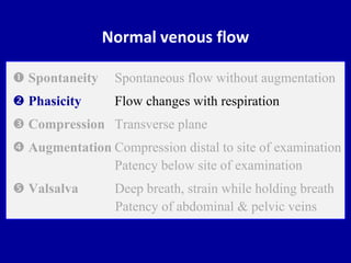
Measurements of phasic blood flow in the inferior and superior venae cavae and umbilical vein of fetal sheep gestational age 121 to 140 days were made with chronically implanted electromagnetic. Normal venous flow Spontaneity Spontaneous flow without augmentation Phasicity Flow changes with respiration Compression Transverse plane Augmentation Compression distal to site of examination Patency below site of examination Valsalva Deep breath strain while holding breath Patency of abdominal pelvic veins 5. PARAMETERS OF NORMAL VENOUS FLOW Venous flow.
The internal jugular vein displayed little phasic variation. In duplex ultrasonography blood flow in standard vein is spontaneous phasic wi Posted on February 28 2013 by admin In duplex ultrasonography blood flow in ordinary vein is spontaneous phasic with respiration and may be augmented by guide stress. Spontaneous non-phasic flow B.
Other types of venous thrombosis such as intra-abdominal and intracranial are discussed in separate articles. The umbilical vein remains intact throughout life but without blood flow. Flow is variable in the hepatic vein.
Minimally phasic continuous Doppler signals in portal splenic and mesenteric veins. Vein is compressible C. When performing venous sonography of a unilateral upper extremity examination.
The breathing-related intra-abdominal pressure changes lead to respiratory fluctuation of venous flow with faster flow during expiration due to lower intraabdominal pressure upward movement of diaphragm and slower flow during inspiration due to higher intraabdominal pressure downward movement of diaphragm. Spontaneous recanalization with blood flow in the umbilical vein may occur in portal hypertension 5 6. Spontaneous pulsatile flow Patient positioning for a venous PPG refill study should be.
Phasic bi-directional pulsatile Doppler signals in IVC renal and hepatic veins. Vian and axillary veins. Respi-ratoryphasicity dominated theflowpatternin nine Fig.
EIV indicates external iliac vein. The upper limb veins displayed weak inspiratory phasicity. Is unidirectional without retrograde flow.
A venous Doppler sonographic examination was considered normal if spontaneous anterograde phasic flow was present and pulsatile if flow had a cyclic retrograde component. Characteristics of spontaneous phasic contractions of the rat portal vein. PSS otherwise known as thoracic outlet syndrome and previously known as effort thrombosis is an uncommon cause of DVT of the subclavian vein most commonly seen as a consequence of chronic compression of the subclavian vein at the level of the thoracic outlet 45Primary axillarysubclavian vein thrombosis was first described by.
A Spontaneous and respirophasic flow with normal response to an augmentation maneuver and aliasing of the pulsed Doppler signal arrow. Supine with head elevated B. B Monophasic venous flow suggesting venous obstruction proximal to this segment.
Blood flow in veins should be _____ and _____ nonspontaneous and nonphasic spontaneous and nonphasic spontaneous and phasic nonspontaneous and phasic. Sometimes a minimal physiological cyclic retrograde flow at the end of the inspiration phase is present a. During inspiration there is minimal flow fluctuation in the portal vein.
Venous flow dynamics differ in the upper and lower extremities. Spontaneous anterograde phasic flow was present and pulsatile if flow had a cyclic retrograde compo nent. Deep and superficial veins of the lower extremity.
Respiratory phasic-itywas mild inone moderate infour and marked with inspiratory flow reversal in seven83Cinterobserver agreement. Some describe scanning the superficial venous system like scanning a plate of spaghetti. As shown in Figure 1 the portal vein possessed a spontaneous phasic contractile activity at a frequency of 003006 Hz and the amplitude of the contraction was 250 - 500 mg which is consistent with previous findings Funaki Bohr 1964.
VENOUS FLOW SPONTANEOUS PHASIC FLOW Venous flow responds to respiration. It is very good at detecting the location of thrombus. Greater Lessor Small Perforators.
Phase polarity was reversed in the lower limbs with near flow stoppage during inspiration. The Doppler waveform is anterograde usually phasic and spontaneous well modulated by breathing. Requires little assistance from the patient.
Surgical reopening of the umbilical vein has been used to create portosystemic shunting in patients with portal hypertension 1 -4. Spontaneous phasic flow C. An advantage of venous plethysmography is.
Deep vein thrombosis DVT most commonly occurs in the lower limbs however are not uncommon in the upper limb and neck veins. Continuous venous flow B. These observations conflict with the current notions of venous flow phasicity which are based on push-pull pressure changes in the abdominal and thoracic.
A prospective controlled study was undertaken to determine how peripheral vascular disease PVD influences flow in the deep veins of the legMethods. Before the development of sonography the clinical diagnosis of lower extremity thrombus was confirmed by venography using invasive injection of contrast material into the lower leg vein. In group 2 36 had pulsatile waveforms and 43 tricuspid regurgitation.
Normal vein Doppler waveform. The pulsed Doppler spectral waveform in a normal lower limb vein exhibits spontaneous and respiro- phasic flow pattern Figure 2. Supine with legs externally rotated D.
Normal lower extremity venous sonography demonstrates spontaneous and phasic flow whereas upper extremity venous flow dynamics are pulsatile due to the proximity of the heart. Eighty-nine patients with peripheral vascular disease and 35 age-matched control subjects were studied. The popliteal vein diameter and flow velocity were measured at rest by means of color duplex.

Spectral Doppler Waveform Analysis Of The Lower Limb Veins Spontaneous Download Scientific Diagram

Spectral Doppler Waveform Analysis Of The External Iliac Vein Eiv Download Scientific Diagram
Doppler Ultrasound Of Normal Venous Flow


Comments
Post a Comment